Breaking News
- Quick Links
- About Us
- Contact Us
- E-Paper

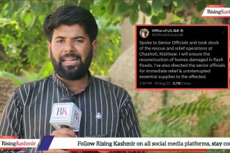
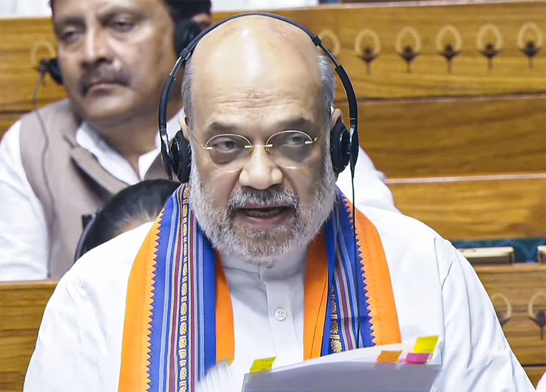
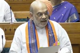
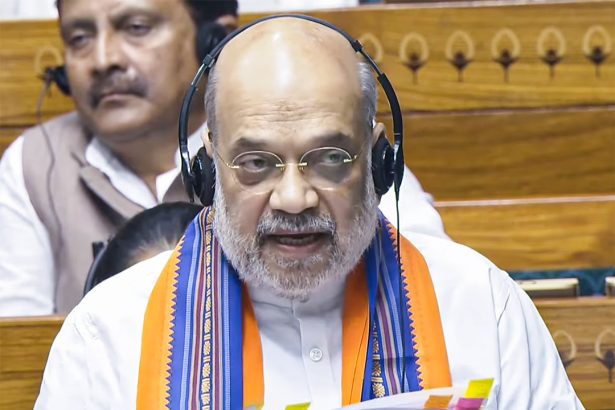
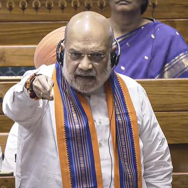
Union Home Minister Amit Shah on Wednesday called on the nation to decide whether it is appropriate for a minister, Chief Minister, or Prime Minister to run the government while…
•Proposed constitutional amendment to disqualify leaders under prolonged custody •New Bills seek to end practice of leaders governing from jail • Disqualification clause to apply uniformly across states & UTs…
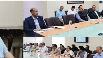


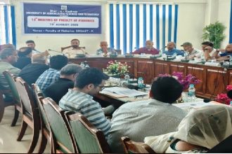
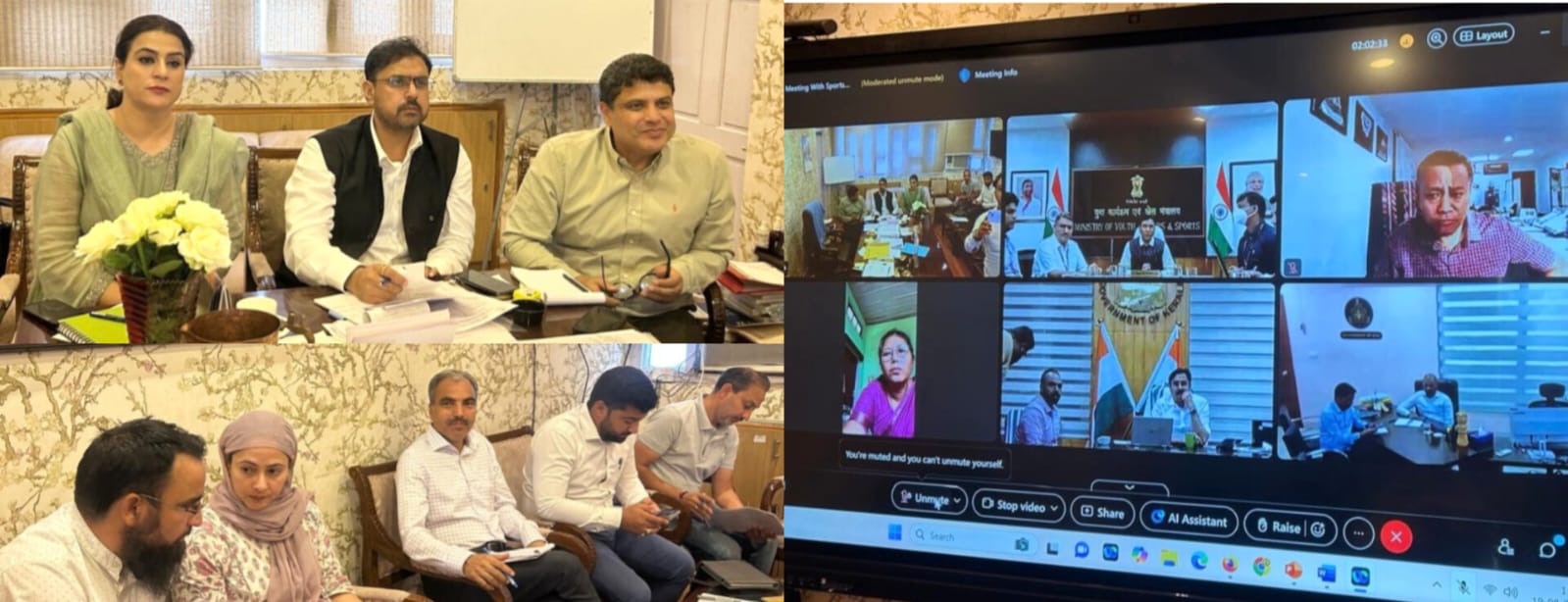
Srinagar, Aug 19: The 12th meeting of the Faculty of Fisheries, Rangil,…

Leh, Aug 20: The Mahabodhi International Meditation Centre (MIMC) Leh, in association…

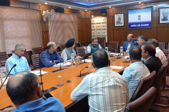
- Geoportals for climate, hazard mapping - Satellite-backed planning tools for Leh,…

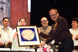
H&H Dept sanctions new units, invests in skill dev to empower artisans,…
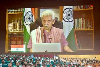
Calls for fostering innovation, critical thinking among youth

We agreed to consider genuine demands of drivers: CEO JSCL

Assure speedy relief, restoration of services
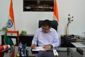
Poonch, Aug 20: Deputy Commissioner Poonch, Ashok Kumar Sharma, Wednesday chaired a…
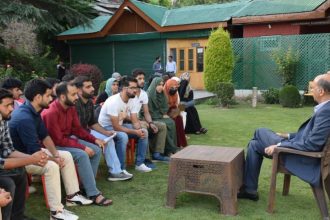
After years of flying across the Indian subcontinent, commercial pilot Captain TanviRaina is charting a new course, this time, through Kashmir’s workshops, drawing rooms, and courtyards. Raina, who continues to…


Over the years there has been the development of a trend to restrain research to conferences and academic discussions. Conventional…
New Delhi, Aug 20: Union Minister Amit Shah’s moving of three Bills…