TOMOGRAPHY (CBCT) AMONG DENTISTS
A good diagnosis is the key to expect the best treatment outcome for patients. Often history and clinical examination are insufficient in assisting dental health professionals in arriving at an appropriate diagnosis. Radiology is diagnostic adjunct in the holistic management of patients. Ever since the ‘dental X ray pioneers’ took the first radiographs in early 1896, radiology has become an integral component in the assessment of patients.
Two-dimensional radiographic techniques commonly used in dental radiology are extraoral radiographs and intraoral radiographs such as panoramic radiographs and cephalometric radiographs. In intraoral radiography, the image receptor is placed inside the patient’s mouth which is used to achieve the desired clinical view, i.e. bite-wing, periapical or occlusal radiographs.
Cross contamination due to intraoral sensor placement and inability to place image receptors in cases of reduced mouth opening are some of the disadvantages of 2-D imaging. Cephalometric radiographs provide low spatial resolution due to overlapping of complex osseous structures. Panoramic images are often obscured by artifacts especially in the anterior region. There is inability to assess width and height of the alveolar bone.
Radiographic evaluation and diagnosis has undergone enormous changes in the last 20 years. New technologies are being developed and are becoming readily available to the medical and dental field. With the expanding array of imaging modalities, dental radiology has played revolutionary role in determining diagnosis, treatment plan and prognostic value.
Although oral health professionals have long relied on 2-D imaging for diagnosis and treatment planning, this technology usually involves radiation exposure. Technological advances, such as digital imaging systems, have greatly increased the level of detailed information available to practitioners while reducing patient exposure to radiation. Today, with a properly prescribed 3-D acquisition, practitioners have acquired the capacity to collect much more data, often with a single acquisition and potentially with much lower radiation exposure to the patient.
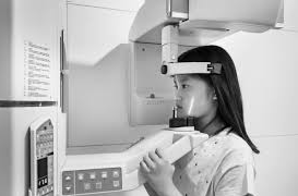
The advent of conical beam computed tomography (CBCT) caused a radical change in dental and maxillofacial radiology. Cone beam computed tomography (CBCT, also referred to as C-arm CT, cone beam volume CT or flat panel CT) is a medical imaging technique consisting of X-ray computed tomography where the X-rays are divergent, forming a cone. It was first introduced into the European market in 1996 and into the United States market in 2001. This type of CT scanner utilizes specific technology to generate three-dimensional (3-D) images of dental structures, soft tissues, nerve paths, and bone in the craniofacial region in a single scan.
Compared to conventional CT scanners, CBCT devices are less expensive and space-efficient, have fast scan time, and confine the beam to the head and neck, reduce radiation doses and have interactive viewing modes that offer maxillofacial imaging and multiplanar reformation, making them more appropriate for use in dental offices.
Generally, CBCT can be categorized into high-volume, medium-volume, and limited units depending on the size of their “field of view”. The cone-beam CT images provide more accurate treatment planning. With cone beam CT, an X-ray cone-shaped beam is moved around the patient to produce a large number of images as views or slices, and it produces high-quality images. One of the advantages of this technology is that it can provide sub-millimeter resolution in terms of images. These images are also of high diagnostic quality.
CBCT is a popular choice amongst dental professionals due to its short scanning time. It takes only 10-70 seconds to finish the acquisition. In addition, it was reported that radiation doses were 15 times lower. Increased use of this system can help dental clinicians receive imaging modalities with the ability to provide a three-dimensional (3-D) representation of a patient’s maxillofacial skeleton. The CBCT performs this function with minimal distortion and is a really useful system for many dentistry professionals.
On the other hand, the information obtained through CBCT imaging also requires a significant level of interpretive expertise. This means that the untrained clinician will likely have a substantial error rate in interpreting CBCT images causing a high percentage of undiagnosed or falsely positive diagnoses.
A recent online survey of active members of the American Association of Endodontists (AAE) in the United States and Canada revealed a significant increase in the use of CBCT, which showed that 34.2% of the 3,844 respondents used CBCT in their clinical practice. The investigation concluded that CBCT was most frequently used in the diagnosis of pathologies as a preparation for endodontic therapy or endodontic surgery, and as a supplement to the diagnosis of trauma-related injuries. CBCT, like every technology, has known limitations. The patient’s history and clinical examination must justify the use of CBCT by demonstrating that the benefits to the patient outweigh the potential risks. Clinicians should only use CBCT when conventional dental radiography or other imaging methods are insufficient to adequately address the need for imaging. There are also many manufacturers and designs of CBCT equipment available.
In India, CBCT has started gaining popularity as preferred imaging modality by the dental practitioners in recent times. There is also a lack of any standardized training modules on CBCT in India. Though some workshops are organized sporadically, but there is no standard curriculum or protocol for the same. The current status of awareness and knowledge regarding CBCT and its indications in dentistry is not known precisely amongst the dental practitioners.
Hence, this study was designed to analyze the current status of the knowledge of the dental students and dental practitioner in Delhi NCR region toward the use of CBCT.
(The Author is III – year post graduate student, oral medicine and radiology at Shree Bankey Bihari Dental College)


