Traditional spinal surgeries like Laminectomy and Lumbar fusion generally involve scars with complete exposure to the patient’s anatomy which requires a high level of expertise to treat a patient’s spinal deformity. But with the recent advancements in technology, Spinal Navigation Technology has completely changed the methods of treating spine ailments. It is now used by neurosurgeons to perform delicate and complex spinal surgeries with low level of risk involved and faster recovery. These image-based technological advancements have made complex procedures easier for spine surgeons as now they are able to operate with better visualization and more accuracy than before.
Spinal Navigation Technology
Navigation System Camera’s track a patient’s anatomy and instruments which produces an image for the surgeon to perform the surgery. This specialized software creates a virtual, 3-D model of the patient’s spine, like a blueprint to help guide the surgeon. The surgeon uses this model to plan the details of the surgery including number, size and location of implants. The surgeon is then able to see and track the position of these surgical instruments and implants in real-time in relation to the patient’s true anatomy. Spine Navigation Technology used for complex surgeries provides faster, more precise and less invasive spinal procedures with less exposure to radiation.
Advantages of Spinal Navigation Technology
Minimally invasive surgery combined with Spinal Navigation Technology provides a less invasive, faster and easier procedure for the patients. The highly effective navigation system helps surgeons perform the most complex spine surgeries. A surgeon may require multiple x-ray images throughout the surgery to guarantee the placement of the implants, while with Spinal Navigation Technology; the need for multiple x-ray images is eliminated as the surgeon can see what is happening right on their screen. Image-guidance plays a major role in making the surgeon’s job easier and saves valuable time. Besides, image-guidance can play a major role in increasing a surgeon’s confidence in difficult cases, especially while performing revision surgeries where the position of the patient’s anatomy may be different from before.
Who can be benefitted by this advanced technological system?
Image-guidance technology helps people of all age groups and can be used in all types of spinal surgeries. Spinal fusion surgeries relieving pain from injury, degenerative disk disease, spinal curvatures or arthritis are currently the most common image navigated surgeries. People who undergo spinal fusion surgeries will experience less pain and will experience an improvement in performing their daily chores.
How is a Navigation Assisted Surgery performed?
Before the surgery, the patient will undergo a pre-operative CT scan inside the OT. These images are then downloaded into the navigation computer to assist the surgeon. The advanced high-tech software then uses these images to build a 3-D model of the spine. The surgeon then uses Smart Instruments in order to match pre-defined points on the 3-D computer model to the patient’s true anatomy. This process is known as registration.
Once this process is completed, the navigation camera starts tracking the movement and position of these smart instruments in the surgical field. The real-time images of these instruments are then displayed on the 3-D model. The monitor shows the exact position of these instruments which helps in surgical precision. Potential damage to surrounding tissue and structures is out of the question because of this image-guidance technology. In spinal fusions, a 3-D model is created to plan the position, length and diameter of pedicle screws, and then the navigation instruments are used to ensure correct positioning of the screw implants. This procedure greatly reduces the risk involved in such surgeries.
Surgical Navigation can’t replace the skill of the surgeon but can help assist in typical complex surgeries with real-time guidance where visibility with the human eye is sometimes challenging.
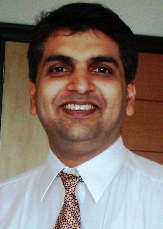
(The Author is Consultant Spine Surgeon at Mumbai Spine Scoliosis & Disc Replacement Centre Bombay Hospital, Mumbai)


